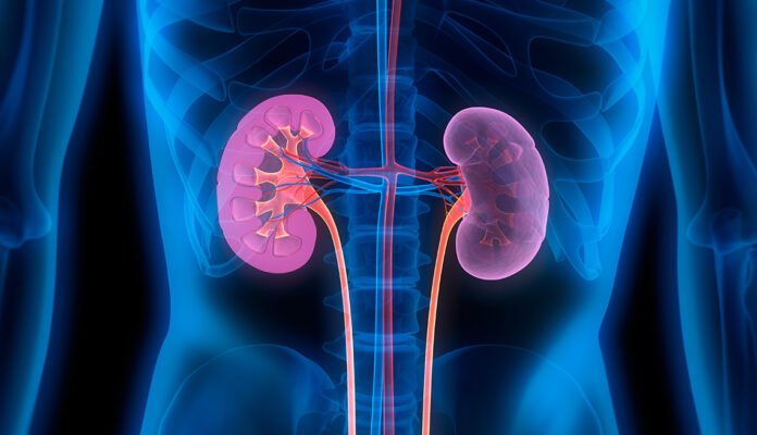
Kidneys are two ruddy earthy colored bean-formed organs found in vertebrates. They are situated on the left and right sides in the retroperitoneal space, and are around 12 centimeters long in grown-up people. They get blood from matched renal corridors; Blood exits in matched renal veins. Every kidney is associated with a ureter, a cylinder that conveys discharged pee to the bladder.
The kidney takes part in the control of different body liquid volumes, liquid assimilation, corrosive base equilibrium, different electrolyte focuses, and evacuation of poisons. Filtration happens in the glomerulus: a fifth of the volume of blood entering the kidneys is sifted. Instances of reabsorbed substances are sans solute water, sodium, bicarbonate, glucose, and amino acids. Instances of emitted substances are hydrogen, ammonium, potassium, and uric corrosive. The nephron is the primary and useful unit of the kidney. Every grown-up human kidney contains around 1 million nephrons, while a rodent kidney has something like 12,500 nephrons. The kidneys likewise work freely of the nephron. For instance, they convert an antecedent of vitamin D into its dynamic structure, calcitriol; And incorporate the chemicals erythropoietin and renin. Follow prozgo for more updates.
Structure
In people, the kidneys are found high in the stomach pit, one on each side of the spine, and in a retroperitoneal position at a marginally diagonal point. The deviation inside the stomach hole, because of the place of the liver, typically makes the right kidney be somewhat lower and more modest than the left, and set marginally higher in the center than the left kidney. The left kidney is around at the vertebral level from T12 to L3, and the right is somewhat beneath. The right kidney sits just beneath the stomach and behind the liver. The left kidney sits underneath the stomach and behind the spleen. Over every kidney is an adrenal organ. The upper pieces of the kidney are to some degree safeguarded by the eleventh and twelfth ribs. Every kidney, with its adrenal organ, is encircled by two layers of fat: the perirenal fat present between the renal belt and the renal container, and the pararenal fat better than the renal sash.
Gross life structures
The practical substance, or parenchyma, of the kidney is partitioned into two significant designs: the external renal cortex and the inward renal medulla. By and large, these designs take the state of eight to 18 cone-formed renal projections, every one of which contains the renal cortex encompassing a piece of the medulla called the renal pyramid. Between the renal pyramids are projections of the cortex called renal sections. The nephrons, the pee creating practical designs of the kidney, range the cortex and medulla. The underlying separating some portion of the nephron is the renal cell, which is situated in the cortex. This is trailed by a renal tubule that runs profound into the medullary pyramid from the cortex. Part of the renal cortex, a medullary beam is an assortment of renal tubules that channel into a solitary gathering conduit.
The tip of each pyramid, or papilla, discharges pee into a little calyx; The minor celiac exhausts into the major celiac, and the major celiac purges into the renal pelvis. It turns into the ureter. In the hilum, the ureter and renal vein leave the kidney and the renal supply route enters. Lymphatic tissue alongside hilar fat and lymph hubs encompass these designs. Hilar fat is adjoining with a fat-filled hole called the renal sinus. The renal sinuses all in all contain the renal pelvis and the calyx and separate these designs from the renal medullary tissue. You must also know about what is cell specilisation.
Blood supply
The kidneys get blood from the renal courses, the left and right, which branch straightforwardly from the stomach aorta. Notwithstanding their moderately little size, the kidneys get around 20% of heart yield. Each renal conduit branches into segmental supply routes, which further separation into interlobar courses, which enter the renal container and reach out through the renal sections between the renal pyramids. The interlobar conduits then, at that point, supply blood to the arcuate corridors going through the boundary of the cortex and medulla. Each arcuate conduit supplies a few interlobular corridors that feed into the afferent arterioles providing the glomeruli.
Blood channels from the kidney, in the long run into the second rate vena cava. After filtration happens, the blood travels through a little organization of more modest veins (veins) that unite into interlobular veins. Similarly as with blood vessel circulation, the veins follow a similar example: the interlobular arcuate veins give blood and afterward return to the interlobar veins, which structure the renal vein leaving the kidney.

